Introduction
The two procedures most often performed for the correction of pectus excavatum are the open method, the “highly-modified Ravitch procedure,” and the minimally invasive “Nuss procedure,” first described by Dr. Ravitch in 1949 and Dr. Donald Nuss in 1998, respectively.
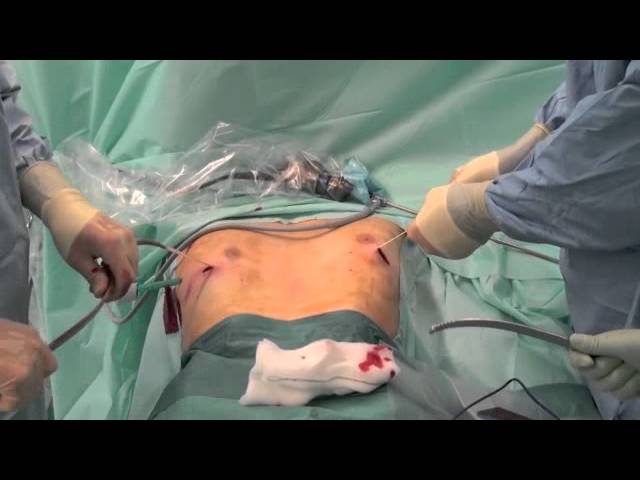
Nuss Technique
The Nuss procedure is considered minimally invasive because it does not require sternotomy or resection of costal cartilages as is necessary in the Ravitch procedure, but instead only requires a few lateral incisions. Briefly, prior to the insertion of the bar, a transthoracic, substernal tunnel travelling anterior to the heart is created using an introducer under thoracosopic guidance. Frantz Then, the arc-shaped bar is inserted with the convexity posterior and then rotated such that the convexity is anterior, displacing the sternum anteriorly. The bar is often fixed using stabilizers or sutures to maintain its position for several years until the cartilage has remodeled and the bar can be removed. The ideal material for the bar is surgical steel rather than titanium because the steel bars have elastic recoil in addition to adequate strength to support the chest in the corrected position if the child sustains trauma.Nuss The bar should be long enough to accommodate growth for two years.Nuss
The standard approach to the minimally invasive technique, including description of patient positioning, thoracoscopic technique, skin incisions, tunneling, sternal elevation, and bar stabilization have been described in detail elsewhere.Nuss
Patients with certain comorbidities or anatomic variants will require special consideration. Older patients, due to the larger volume of their thoracic cage, often require two support bars. Those with Marfan’s syndrome or other connective tissue disorders have softer bones and have better outcomes using two bars to distribute the pressures over a wider area.Nuss Those with an allergy to nickel may receive titanium bars to avoid this complication.
Modifications to the standard Nuss procedure
Displacement of the support bar has been a common complication of the Nuss procedure. Without stabilizers, bar displacement was found to occur in up to 15% of cases.Nuss Many modifications have been described involving methods of fixation with stabilizers to prevent bar dislocation.Clark The development of stabilizers has decreased the incidence of bar displacement, but the different materials, placement, and design of each type of stabilizer present unique challenges.
Switching from rectangular stabilizers to triangular stabilizers and the placement of a circum-costal wire for additional fixation has been reported to reduce the rate of bar displacement to 5% in one study.Nuss A pericostal suture can be placed around one costal arch, often through the right lateral thoracic incision, and a stabilizer placed on the left side.Hebra
A disadvantage of metal stabilizers is that they often cause chronic pain that requires a second intervention for removal.Pilegaard An alternative, slowly absorbable stabilizer made of L-lactic and glycolic acids (Lactosorb®) was introduced in 2007. In one study, of the 85 patients with absorbable stabilizers placed, three stabilizers broke within 6 weeks of surgery (3.5%) and another three absorbable stabilizers (3.5%) dislocated within 8 weeks of surgery without any connection to physical activity. Pilegaard
There is much concern for increased infections with use of stabilizers of any material. However, it was found that while the stabilizers did increase the incidence of wound infections, the infections were initially aseptic seromas with dermatitis due to pressure damage that then became infected after surgical drainage.Watanabe
The standard technique has been to place stabilizers at the bar edges, but Milanez et al. proposed that placement of two stabilizers in the intermediate region between the bar edges and the mid-point, close to the entry and exit of the bar in the intercostal space, posterior to the muscles, but not close to the cartilage, would be less likely to become displaced. They used stabilizers with central grooves on the posterior surface that allowed for the stabilizer to slide easily, regardless of the bar’s curvature. The disadvantage of this technique is that it requires the additional dissection of the pectoralis major muscles medially to allow the stabilizers to be placed posterior to them. Milanez et al. used this technique in two patients, without any additional morbidity or complication incurred.
The edges of the bar are serrated, and can cause tissue damage and hemorrhage when it is removed, particularly if the patient is not maintained in a symmetrical position throughout removal.Nuss Milanez et al. have introduced a technique in which the serrated edges of the bar are enveloped in a protective film to protect the surrounding tissue during removal. This technique was used for one patient, without any additional morbidity or complication incurred.
To further minimize scarring, the Nuss procedure can be performed using only a right lateral thoracic incision with results comparable in safety and efficacy to the standard Nuss technique.Clark Through a single right lateral thoracic incision and under thoracoscopic guidance, a dissection across the chest in a subcutaneous, pre-sternal, and pre-muscular plane forms a tunnel into which the bar can be placed in the standard position. No statistically significant differences between the study groups in any of the patient, operative, or postoperative care parameters were reported.Clark
Outcomes of the Nuss Procedure
Surgical repair of pectus excavatum with the Nuss procedure has been shown to improve parameters of cardiovascular function such as cardiac stroke volume, cardiac output, forced expiratory volume in the first second of expiration (FEV1), total lung capacity (TLC), TLC (percentage expected), diffusing lung capacity, maximum oxygen consumption (VO2max), respiratory quotient, and O2 pulse (percentage predicted).Chen A study on the end-diastolic right ventricular dimensions and left ventricular ejection fraction of adults using intraoperative transesophageal echocardiography immediately before and after surgical correction of pectus showed that both parameters immediately and significantly increased after surgical correction.Krueger
There is a modest preoperative reduction in vital capacity and total lung capacity, both of which improve after surgical correction.Chen FEV1, the amount of air that can be forcefully exhaled in one second, was improved postoperatively. This indicates that the repaired chest wall allows the extrinsic musculature and diaphragm to facilitate more efficient breathing. The Ravitch and Nuss procedures effect equal improvement in pulmonary function within 1 year, but greater improvements were seen for Nuss patients after bar removal.Chen
Others found that postoperative Nuss procedure patients had no change in lung function
“Postoperatively, patients who have undergone the Nuss procedure typically experience a temporary decrease in forced vital capacity, which resolves with removal of the implanted bar [6]. The likely explanation is that the bar restricts maximal thoracic expansion. In support of this, a study by Castellani et al. [6] showed no significant differences in preoperative and postoperative lung function.”Dean
The measured improvements in cardiopulmonary function may be confounded by the post-operative cardiorespiratory rehabilitation that patients receive, improved optimism toward exercise goals, and increased social integration.
The Single Step Questionnaire (SSQ), is a questionnaire designed to assess the impact of surgical correction of PE on patient’s quality of life and psychosocial well-being. Administration of the questionnaire to forty patients at 6 months after MIRPE with the bar in place and again to the patients and their parents at a mean of 23 months after bar removal demonstrated a significant improvement in parameters linked to self-perception, self-esteem and social behavior.Metzelder
Nuss Complications
Common complications of the Nuss procedure include pneumothorax, pleural effusion, pain, wound infection, and bar displacement.Vegunta Less common complications include cardiac perforation, aortic perforation, pericarditis, brachial plexus injury, diaphragmatic perforation, and thoracic outlet syndrome.Vegunta The use of thoracoscopy has significantly decreased the incidence of cardiac complications, but perforations have occurred despite this protective measure.Bouchard Left chest thoracoscopy with blunt dissections provides improved mediastinal visualization and a decreased risk of cardiac injury, except in those with severe deformities and a leftward displaced heart.Bouchard The support bar exerts a force that ultimately is absorbed by the spine. If the malformation is asymmetric, the spine may bend from the side of the chest with greater concavity to the side with less concavity, resulting in scoliosis. Interestingly, this remodeling may be advantageous if the side of spinal bowing coincides with the side of anterior wall concavity because the spinal bowing may be corrected by that counterforce.Dean
Ravitch Technique
“In 1949, Ravitch described a technique (open technique) involving dissection of the pectoralis major and rectus abdominis, followed by subperichondrial resection of all deformed cartilages, xiphoid excision, and sternal osteotomy with anterior fixation of the sternum .”Dean Modifications to the Ravitch technique have focused on experimentation with different materials for posterior support of the sternum.
Termed the “Robicsek technique,” a modification of the Ravitch procedure involving the use of DualMesh 2-mm Gore-Tex to provide posterior support of the sternum has been reported on with great results by several authors.Kotoulas, Spartalis Spartalis et al. report the results of a 17 year old patient who underwent this modified Robicsek technique. Their technique is described as follows: the mesh is placed under the sternum and anchored to the lateral tips of all divided costal cartilages to stabilize the chest wall. The previously detached edges of pectoralis major and rectus abdominis muscles were sutured over the anterior sternal surface. The pectoral muscle flaps were secured to the midline while being advanced to provide coverage of the entire sternum. The rectus abdominis muscle flap was joined to the pectoral muscle flaps. The patient had no postoperative complications and had excellent cosmetic results. A high resolution three-dimensional reconstructed computed tomographic scan taken seven years post-operatively confirmed that the mesh was still stably anchored and supporting the sternum in its corrected position. Two main advantages of the Gore-Tex mesh are that it is very resistant to infections and it has two different surfaces: one smooth for minimal tissue attachment and the other textured for tissue ingrowth.Kotoulas The textured surface is positioned adjacent to the posterior sternum and causes creation of adhesions, while the smooth surface faces the pericardium and does not cause adhesions to form. Thus, the authors propose that the material may stay in place permanently whereas at the same time it can be detached easily from the pericardium in the case of an urgent sternotomy.Kotoulas Kotoulas et al., with 10 years experience using the 2-mm Gore-Tex mesh support in 21 young adult patients, reported “excellent cosmetic results, no cases of infections, no cases of recurrence, and no limitations in activity.”
Comparison of Nuss and Ravitch Procedures
Advantages of the Nuss procedure are that it avoids extensive dissection, cartilage resection and osteotomy, which are integral components of the Ravitch technique.Dean It requires a shorter operating time, results in only minimal blood loss (in the 10-30 mL range), and allows for an earlier return to full activity.Nuss It provides superior cosmetic results, which is often an important consideration for patients as cosmesis is often a primary motive for seeking correction.Krasopoulos
Compared to pre-adolescents, adult patients undergoing the Nuss procedure often experienced more intense pain postoperatively, required prolonged postoperative care, and had a higher incidence of bar displacement and recurrence; all of which may be attributable to the increased rigidity of the chest wall in these older patients.de Souza Coelho Outcomes for the Nuss procedure performed on adults were better if two bars and/or lateral stabilizers were used.Dean Patients with asymmetric malformations who were corrected using the Nuss procedure were more likely to be over corrected and develop postoperative pectus carinatum than those with asymmetric malformations who underwent the Ravitch procedure.de Souza Coelho Thus, open repair should be considered for the older patient or the patient with an asymmetric malformation.
Failed Repairs and Recurrence
The recurrence rate after repair of PE is reported to be anywhere from 2% to 37%.Croitoru Recurrences are more common in those with Marfan’s Syndrome, those whose repair was completed at either a very young or an advanced age, those with either too extensive or too little dissection with an open repair, premature removal of the bar in minimally invasive repairs, local infections, or an unsatisfactory primary result.Croitoru Patients with a failed repair or recurrence will often present with the same symptoms they experienced before the primary repair, such as shortness of breath, chest pain, and other similar symptoms that affect their activity level.Croitoru Associated cardiac findings such as cardiac displacement and cardiac compression were often still present.Croitoru In addition, these patients continued to perform poorly on pulmonary function tests (PFTs).Croitoru Patients with a failed Nuss procedure will usually present shortly after the primary surgery with bar displacement will be evident on chest x-ray.Croitoru Patients with a failed Ravitch procedure or a recurrence after such may present immediately after or up to seven years postoperatively.Croitoru Those that present remote from the primary surgery are often those that had a successful repair performed before the pubertal growth spurt but the thoracic cage failed to grow appropriately during puberty.Croitoru
Croitoru, et al., reported excellent results with the minimally invasive Nuss technique for recurrent or failed pectus excavatum repair. Their study demonstrated that the Nuss procedure was superior to the open procedure for a repeat repair. The inherent extensive dissection performed during an open procedure, although technically extrapleural, can produce extensive intrathoracic adhesions. Also, most patients with a previous Ravitch repair will have poor chest wall movement and compliance because of the abnormal ossification of the anterior chest wall secondary to the previous invasive procedure. Repeating the open procedure would necessitate another violation of the perichondrium and periosteum, thus stimulating more ossification, calcification, and subsequent loss of chest wall movement and compliance. For this reason, Croitoru et al., also do not advocate wiring of the pectus bar to the ribs or sternum. In patients undergoing a second repair, “complications were slightly higher than those in primary repairs and included pneumothorax requiring chest tube (14%), hemothorax (8%), pleural effusion requiring drainage (8%), pericarditis (4%), pneumonia (4%), and wound infection (2%). There were no deaths or cardiac perforations. Initial postoperative results were excellent in 70%, good in 28%, and fair in 2%. Late complications of bar shift requiring revision occurred in 8%. Seventeen patients have had bar removals with 9 patients being more than 1 year postremoval. For the 17 patients who are postremoval, excellent results have been maintained in 8 (47%), good in 7 (41%), fair in 1 (6%), and failed in 1 (6%). There have been no recurrences postremoval.”Croitoru Although postoperative cardiology evaluations or repeat CT scans were not performed, symptoms of cardiac compression and displacement were found to be relieved postoperatively. The patients who had a secondary minimally invasive procedure after either a primary Ravitch or Nuss procedure and who returned for follow-up reported resolution of their pre-operative symptoms with 85% reporting increased exercise tolerance. 82% of respondents (23/28) reported satisfaction with their results.Croitoru
Summary
Pectus excavatum is a common, congenital chest wall deformity in which the sternum is depressed. In severe disease, the patient may experience cardiopulmonary dysfunction, most commonly described as exercise intolerance. The minimally invasive Nuss procedure and the modified open Ravitch procedure are both safe and effective techniques for repair of pectus excavatum. The choice of which procedure to perform should be made on an individual basis. Multiple factors such as the patient’s age, comorbidities, severity of the defect, and symmetry of the chest wall should all be considered in pre-operative evaluation. Research is ongoing and surgical techniques are continuously evolving as new modifications are introduced.
